M.A. HANSON RESEARCH
  • Home
  • Research
  • Publications
  • Gallery
  • Blog
  • Contact

Images of Drosophila

 From left to right: 1. Drosophila melanogaster infected by green fluorescent bacteria. 2. Drosophila innubila on a mushroom cap. 3. Drosophila neotestacea on a mushroom cap. 4. Clip art of a D. melanogaster female.

Models of antimicrobial peptides

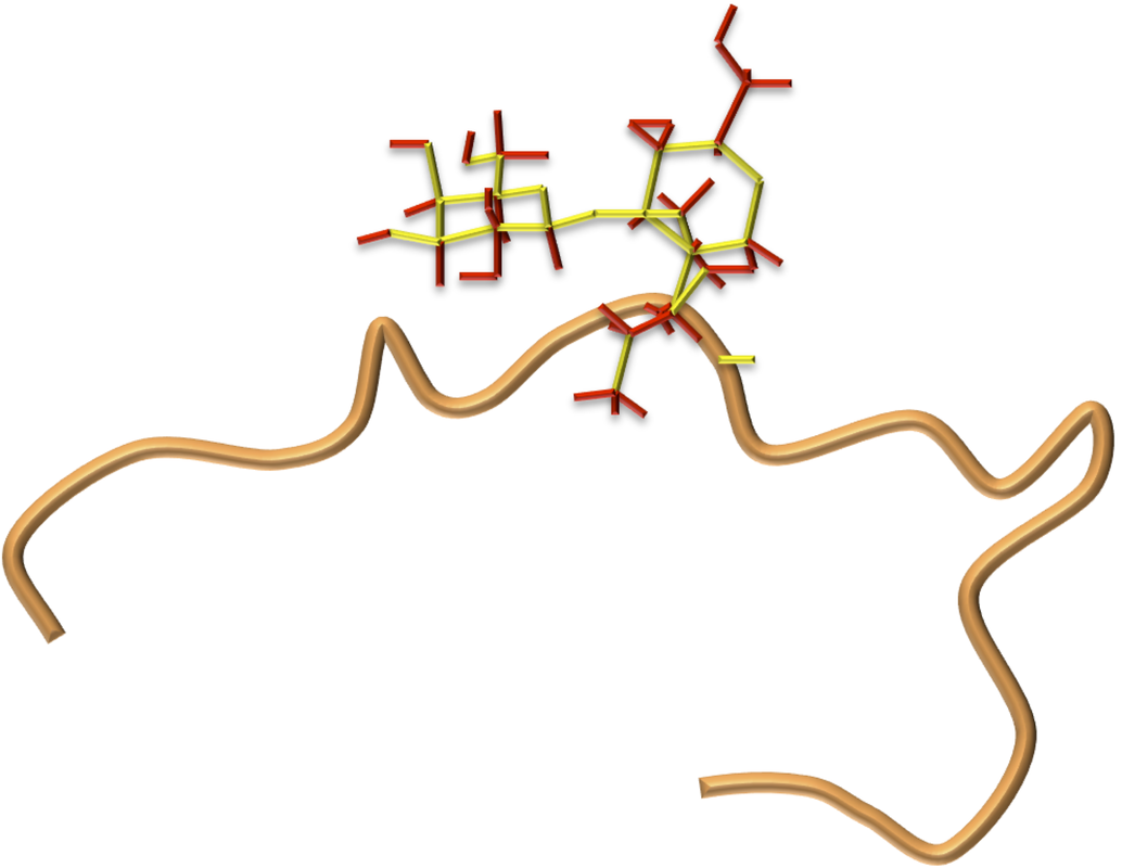
From left to right: 1. 3D model of Drosomycin. 2. 3D model of Drosocin. 3. 3D model of Cecropin. 4. 3D model of the fly Defensin.
All images on this page are free to use under CC-BY-SA 4.0. Please credit "MA Hanson."
Powered by Create your own unique website with customizable templates.
  • Home
  • Research
  • Publications
  • Gallery
  • Blog
  • Contact